Analysis of Protein Expression Profiles
The development of antibody-based technologies has been driven by the low correlation between mRNA presence and protein expression. Antibody Microarray are an extremely useful tool for biomarker study and for a proteomic approach in the study of diseases and/or experimental models. The antibody microarray technology allows to study changes in the expression of a large number of proteins in much less time than would be required with traditional methods and, in a single experiment, allows a complete overview of protein expression levels involved in different pathways.
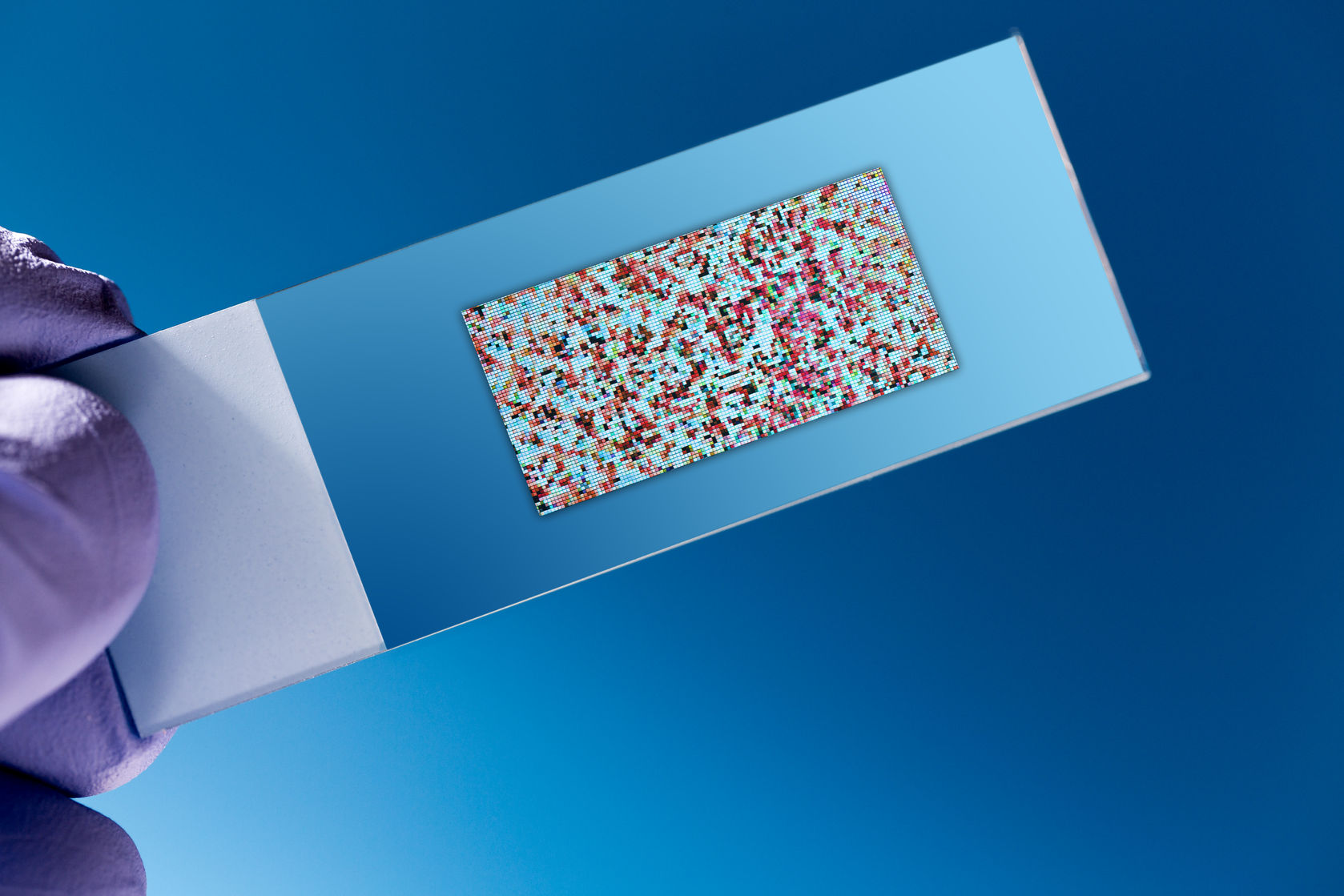
Microgem Lab provides the “Analysis of Protein Expression Profiles” service by using antibody microarray to monitor the binding of proteins labeled in cellular and tissue lysates, serum, plasma.
Workflow of the “One-Color Analysis of Protein Expression Profiles” service.
- Consultancy – A free consultation will be provided for project optimization (number of samples to be analyzed, methodology, costs). During the consultation phase advice will be given on the method of extraction and/or treatment of the sample to be taken. Alternatively, you can take advantage of our protein isolation service.
- Sample Requirements –
- 1 ml of cell culture supernatant with low serum
- 2×10⁷ cell pellet
- 100 mg of tissue
- Cell or tissue lysates at concentration of 2 mg/ml
- 1 ml serum or plasma of human, mouse, rat
- Quality Control of the Sample (QC) – After receiving the samples, our researchers will evaluate the concentration of total proteins (cellular and tissue extracts) by colorimetric assay. The results of this analysis will be sent before proceeding with the next steps of the service.
- Labeling, Hybridization and Reading of the Microarray – The sample (lysate, serum, plasma, supernatant) will be dialyzed, labeled with biotin and hybridized on microarrays on which hundreds of specific antibodies are detected. On each microarray, positive and negative controls are also detected for auto track and cross-hybridization. The antibody-protein complex will be labeled with streptavidin conjugated with a fluorophore. After washing, the microarray will be analyzed by scanner and and the image analysis will be performed.
- Data Analysis – The median of the fluorescence values obtained for each spot will be subtracted from the background value. Subsequently, data obtained from independent microarrays can be compared by normalization using a reference array. A signal intensity comparison will be used to determine differences in expression levels of each protein between sample groups.
- Final Report – At the end of the project, a final report will be provided including the results of the various stages of project development. The results presented in the report can be discussed with the scientific team.
Schede del servizio
- ANTIBODY ARRAY 90 PROTEINE TARGET RATTO Scarica il file .pdf
- ANTIBODY ARRAY 182 ADIPOCHINE UOMO Scarica il file .pdf
- ANTIBODY ARRAY 308 PROTEINE TARGET TOPO Scarica il file .pdf
- ANTIBODY ARRAY 493 PROTEINE TARGET UOMO Scarica il file .pdf
- ANTIBODY ARRAY 507 PROTEINE TARGET UOMO Scarica il file .pdf
- ANTIBODY ARRAY 1000 PROTEINE TARGET UOMO Scarica il file .pdf